How we approached coronary calcium scoring
By Naveen K, Co-Founder and Chief of AI ·
Medically reviewed by Dr. Vishwanath R S, MD, CIIP
Coronary artery calcium is one of the strongest predictors of a future cardiac event. The score is simple to define and tedious to produce, and that combination is where good software earns its place. Today, calcium scoring in most centres happens on a separate workstation, or on an AI server that sends back a screenshot, which means an extra step, an extra system and a result the report cannot easily use.
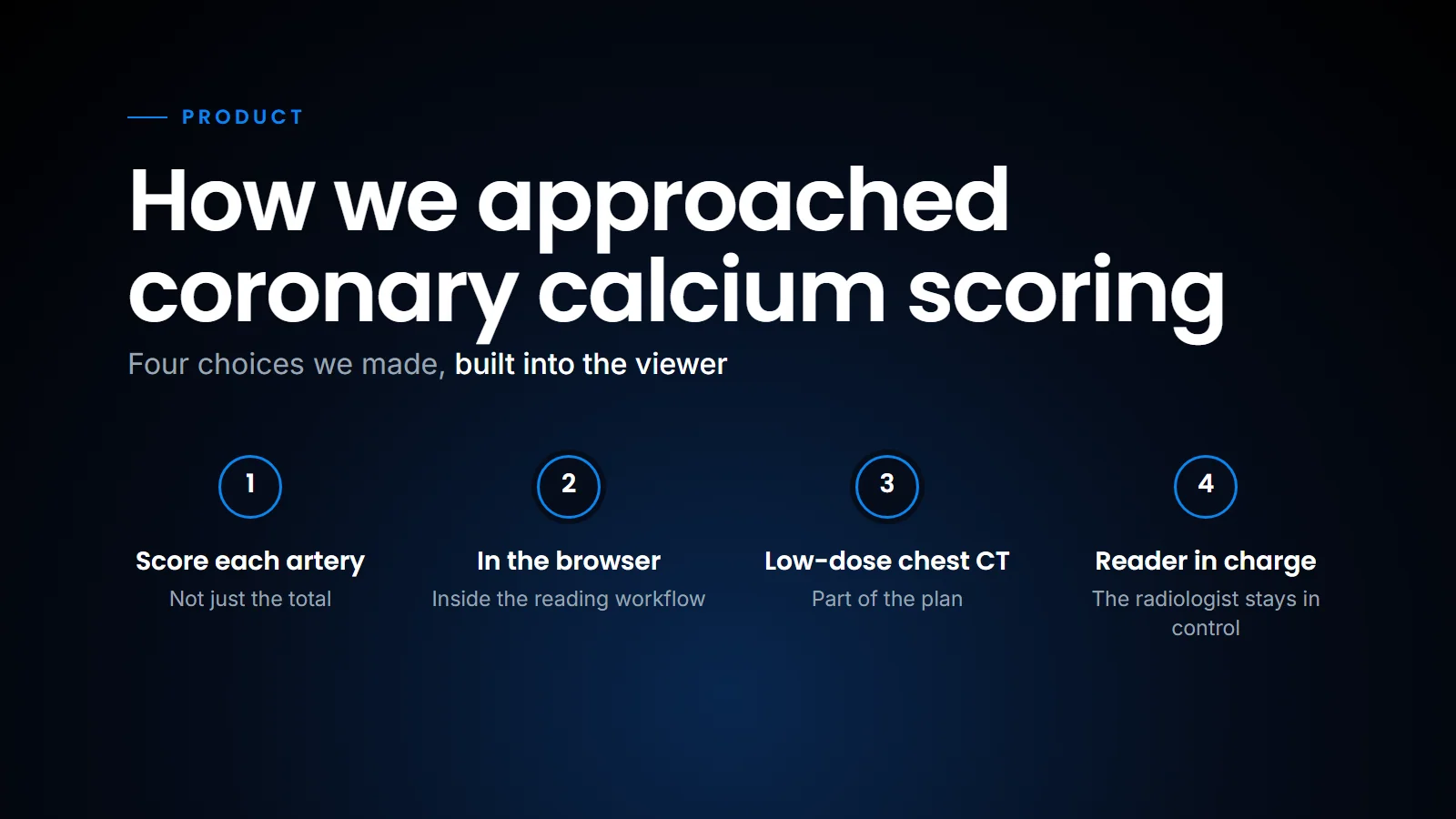
This post covers the method we follow and the four choices we made building it into the SOMATIQ viewer.
Let's start with the method itself.
The Agatston method in one paragraph
Agatston scoring works slice by slice on a non-contrast CT. Any coronary lesion of 130 HU or more, with an area of at least 1 mm², counts. Each lesion's area is multiplied by a weight set by its peak density, and the slice scores add up to the total. The score assumes a standard non-contrast protocol (on modern CT, typically 3 mm slices at 120 kVp), so scores from other protocols, including many low-dose chest CTs, are estimates rather than directly comparable figures.
How the Agatston score is built, from calcium voxels to a risk band.
| Peak density (HU) | Weight |
|---|---|
| 130–199 | 1 |
| 200–299 | 2 |
| 300–399 | 3 |
| 400 and above | 4 |
The total then falls into the familiar bands: 0, 1–99, 100–399 and 400 or more.
Choice 1: score each artery, not just the total
A total of 300 in one vessel is not the same patient as 300 spread across four. We score the left main, LAD, circumflex and RCA separately, then add them up. The cardiologist sees where the calcium is, not just how much there is. The advantage is clinical information; the cost is that someone has to decide which artery each lesion belongs to, which brings us to the second choice.
Choice 2: in the browser, inside the reading workflow
Calcium scoring usually lives on a separate workstation or a separate AI server that sends back a screenshot. We put it in the viewer the radiologist already has open, the same zero-footprint viewer they read every other study in.
The viewer highlights every candidate at or above 130 HU. The reader confirms which ones are coronary and which artery each belongs to. The per-artery and total scores update as they go. Nothing leaves the browser to be computed elsewhere.
This is the same pattern as our PET/CT tools, where a threshold inside a radiologist-placed region gives a 3D lesion segment (see DICOM SEG and PET/CT lesion segmentation and Reading PET/CT in the browser). A fixed, visible threshold does the tedious part, and the radiologist does the judging.
Choice 3: low-dose chest CT matters
The classic score is defined on ECG-gated cardiac CT. In India, far more patients get a low-dose chest CT for lung screening or follow-up than a gated calcium scan. Their coronary calcium is visible in those images and is often not reported.
Non-gated scans blur with heart motion, so the same calcium can score differently than on a gated scan. We treat these results as an opportunistic finding, report them as such, and recommend a dedicated scan where it changes management. A short template line for coronary calcium on every plain chest CT makes this routine rather than a memory test (we discuss templates in Structured reporting). With the platform now running across more than 100 imaging centres, the number of chest CTs where this applies is large; we will look at other opportunistic findings on chest CT in a later post.
Choice 4: keep the reader in charge
The threshold and the weights are fixed by the Agatston method; the risk bands are a reporting convention, and different systems draw them differently (CAC-DRS, for example, uses 1–99, 100–299 and 300 or more), so the report should say which one it uses. What needs judgement is what counts as coronary: valve and aortic calcium, stents, noise and the boundaries between arteries. That judgement stays with the radiologist, and the software makes it fast. It is the same position we took in AI is with the radiologist: the tool proposes, the radiologist decides and signs.
In conclusion, the Agatston method has not changed in decades; what has changed is where it can run. Putting it in the reading viewer, per artery, and applying it to the low-dose chest CTs that Indian centres already do in large numbers turns a separate test into a line in a report the radiologist is already writing.
Related reading
- DICOM SR: AI findings a report can read — carrying a score into the report as data.
- Reading PET/CT in the browser — the other quantitative tool in the same viewer.
- What is a zero-footprint DICOM viewer — why nothing needs installing.
FAQ
What is an Agatston score?
A coronary calcium score from non-contrast CT: lesion area times a density weight, summed over all slices and lesions.
Why score per artery?
The distribution of calcium adds information beyond the total, especially for left main and proximal LAD disease.
Can calcium be scored on a low-dose chest CT?
It can be detected and estimated. Gated cardiac CT remains the reference for a formal score.
Does it need a separate workstation?
No. In SOMATIQ it runs in the browser viewer.
Ready to see your workflow on one platform?
We will walk you through the whole workflow, from registration to signed report.