The collaborative DICOM viewer
Radiology is often described as a lonely specialty. One person, a dark room, a worklist. But the hard cases have never been read alone.
A resident calls the consultant over. A general radiologist sends a study to a neuroradiologist friend at 11 p.m. with "can you have a quick look?" A surgeon walks into the reporting room and asks to be shown exactly where the collection is. A tumour board puts twelve people in front of one screen.
All of this is collaboration. Most of it, in Indian centres, happens over WhatsApp screenshots, phone calls and CDs. These work, after a fashion, and they cost nothing to set up. Their disadvantage is that with a screenshot or a phone call the person on the other end never sees the actual study, while a CD carries the full study but takes days to arrive and needs a viewer on the other side; either way, the patient's images end up in places nobody controls. So, let's look at what a collaborative viewer changes, where it matters most, and what it does not replace.
What we heard at IRIA 2026
When we showed our platform at IRIA 2026 in Hyderabad, the collaborative viewer was especially well received by academic radiologists. What they valued was the ability to securely share their viewer and collaborate with peers anytime, anywhere. (We wrote up the wider conversations in What radiologists told us at IRIA 2026.)
That reaction made sense to us. Academic radiologists teach, take second-opinion requests and sit in on multidisciplinary meetings every week. They feel the gap between what they need and what their tools allow more than most.
The problem with screenshots
A screenshot of a CT slice is a photograph of one moment in a three-dimensional study. The person receiving it cannot scroll. Cannot change the window. Cannot look at the slice above, or reformat in another plane, or compare with the prior.
So the conversation goes like this. "Can you send the slice above?" "Can you send it in lung window?" "Is there a prior?" Five messages later, both radiologists have spent more time than if they had looked at the study together for two minutes.
Screenshots also travel. They sit in phone galleries and group chats, often with the patient's name visible in the corner. That is a privacy problem, and under India's DPDP framework it is not one a centre should be casual about. For what the law actually requires, read the Act and Rules on the MeitY site and talk to your counsel; our guide to the DPDP Act and your radiology data walks through the parts that touch imaging.
What a collaborative viewer changes
The idea is simple. Instead of sending a picture of the study, you share the study itself, in a viewer, with the person you want to discuss it with.
They see the real images, at full quality. They can scroll, window and reformat. You can both look at the same thing and talk about it. And access is controlled, so the study is shared with a specific person for a specific purpose, not dropped into a chat.
Our viewer runs in the browser, built in-house, with rendering on each user's own machine. It supports MPR, MIP and 3D. Because there is nothing to install (see what a zero-footprint viewer is), the person you share with does not need a workstation or a licence on their side to see the study properly.
Four places it matters
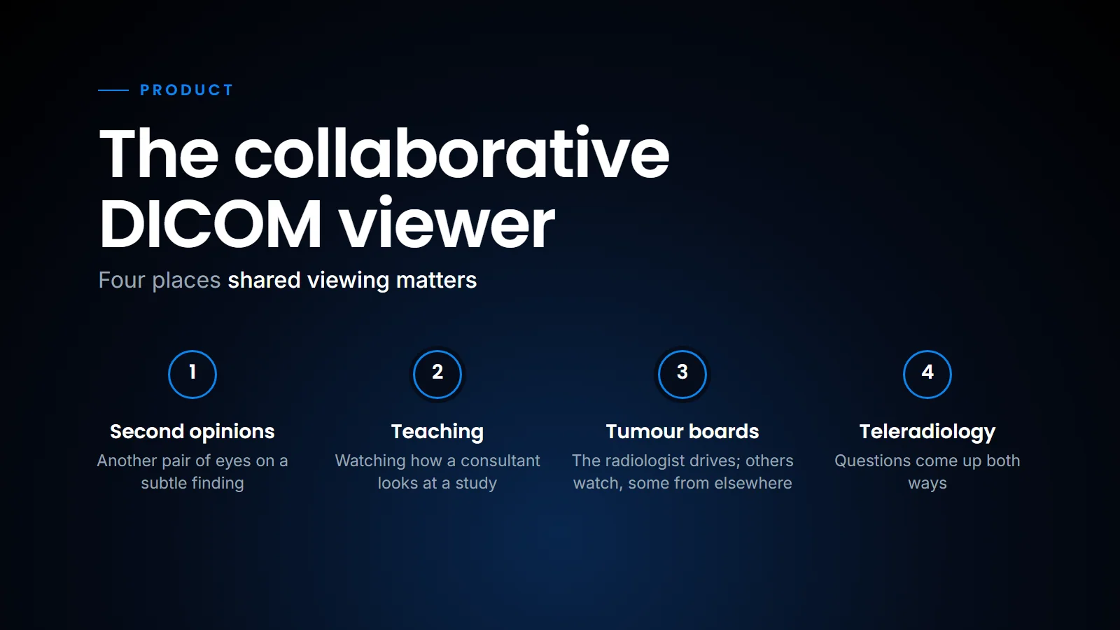
In practice, shared viewing earns its place in four situations.
- Second opinions. The most common use, and the most informal. A radiologist wants another pair of eyes on a subtle finding. Today that means a phone call and a screenshot, or waiting until a colleague is in the same building. With a shared viewer, the colleague looks at the actual study, from wherever they are. They can form their own view of it rather than reacting to whatever slice was chosen for them. That is a better second opinion. (Formal peer review is a different process; on our platform it has its own peer review queue.)
- Teaching. Teaching radiology is teaching how to look. A resident learns more from watching a consultant scroll through a study, stop, adjust the window and point, than from any number of static key images. Shared viewing lets that happen when teacher and student are not in the same room. A consultant at one centre can walk residents at another through a case, and the cases can be real ones from the day's work, which are often more instructive than textbook images. The ones worth keeping can go into a teaching archive with case tags, so they can be found again.
- Tumour boards and multidisciplinary meetings. At a tumour board the radiologist is usually the person driving the images while oncologists, surgeons and pathologists watch. Often some of those people are dialling in from elsewhere, looking at a shaky video of someone else's screen. When participants can open the same study in a viewer, the surgeon who wants to see the relationship of the mass to the vessel can look at it in the plane they care about, and the discussion becomes more precise. This matters most for studies like PET/CT, where fusion and lesion volumes are part of the conversation (see Reading PET/CT in the browser).
- Teleradiology. Teleradiology is collaboration by definition. The reading radiologist is in one place, the patient and the referring clinician in another. Questions come up both ways. TelePractice, our teleradiology platform, is built for this kind of work. Whatever the platform, a shared viewer makes those conversations about the actual images rather than about a description of them.
What it does not replace, and what to ask
A shared viewer is a tool for conversation. It does not change who is responsible for the report. The radiologist who signs is still the radiologist who signs.
It also does not replace referring-doctor access. For a clinician who simply wants to see their patient's images and report, a secure study link is usually the right tool. On our platform, centres can send study links to referring doctors through WhatsApp: the link travels on the app everyone already uses, and the images stay in the viewer. Collaboration is for when two people need to look and talk.
If you run a centre with more than one radiologist, or more than one site, ask yourself a few questions.
- How do your radiologists ask each other for help today? If the honest answer is WhatsApp screenshots, you have both a quality gap and a privacy gap.
- Who outside the centre needs to see images regularly? Consultants at a teaching hospital, surgeons, oncologists, a teleradiology partner. Each is a candidate for shared access rather than screenshots or CDs.
- Can your current viewer share a study securely, with a named person, without installing anything? If not, collaboration will keep leaking into informal channels.
- Do you run teaching or case conferences? A shared browser viewer makes it possible to include people who are not in the room.
SOMATIQ is now deployed across more than 100 imaging centres in three countries. The more distributed a network becomes, the more radiology depends on people being able to look at the same images from different places. That is what a collaborative viewer is for: it keeps the hard case a conversation between people looking at the same study, rather than a chain of screenshots.
Related reading
- Sharing scans with referring doctors — secure links versus photos of the monitor.
- What is a zero-footprint DICOM viewer — why nothing needs to be installed on the other side.
- Running a diagnostic chain on one system — one worklist and archive across centres.
- Reading PET/CT in the browser — fusion and segmentation without a workstation.
FAQ
What is a collaborative DICOM viewer?
It is a medical image viewer that lets a radiologist securely share a study, not a screenshot, with a colleague so that both can look at the full images and discuss them. In a browser-based viewer, the person receiving the share does not need to install anything.
Is it safe to share radiology images on WhatsApp?
Sending screenshots with patient details through chat apps creates privacy risks, because the images are stored on phones and can be forwarded. Sharing a secure link or a controlled viewer session avoids attaching images to the chat and lets the centre control and log access; images are still displayed on the recipient's device, so download and caching settings matter too. For your obligations under India's DPDP Act, consult the Act and Rules and talk to your counsel.
How does a collaborative viewer help tumour boards?
It allows participants to open and look at the same study themselves, in the planes and windows they care about, instead of watching a screen share of someone else's viewer. That makes discussions about anatomy and extent more precise.
Does sharing a viewer change who is responsible for the report?
No. Collaboration helps radiologists discuss a case, but the radiologist who signs the report remains responsible for it.
Ready to see your workflow on one platform?
We will walk you through the whole workflow, from registration to signed report.