Hanging protocols, explained
By SOMATIQ team ·
The term comes from film. Before PACS, a technician or a junior resident would hang the films on the lightbox in the order the radiologist liked: current PA chest on the left, the old one on the right, lateral below. Each consultant had their own preference, and a good technician knew all of them. A hanging protocol is the digital version of that technician. It is a set of rules that decides, when a study opens, which series go where on the screen, in what orientation, with what window, and next to which prior.
When it works, nobody notices. When it does not, the radiologist spends the first thirty seconds of every case dragging series into viewports. Over 150 cases a day, that is real time, and it is the kind of small friction that makes people dislike their PACS. So, let's look at what a protocol actually specifies, why protocols break (usually on priors), what the DICOM standard offers, and what a centre can do about it.
What a hanging protocol actually specifies
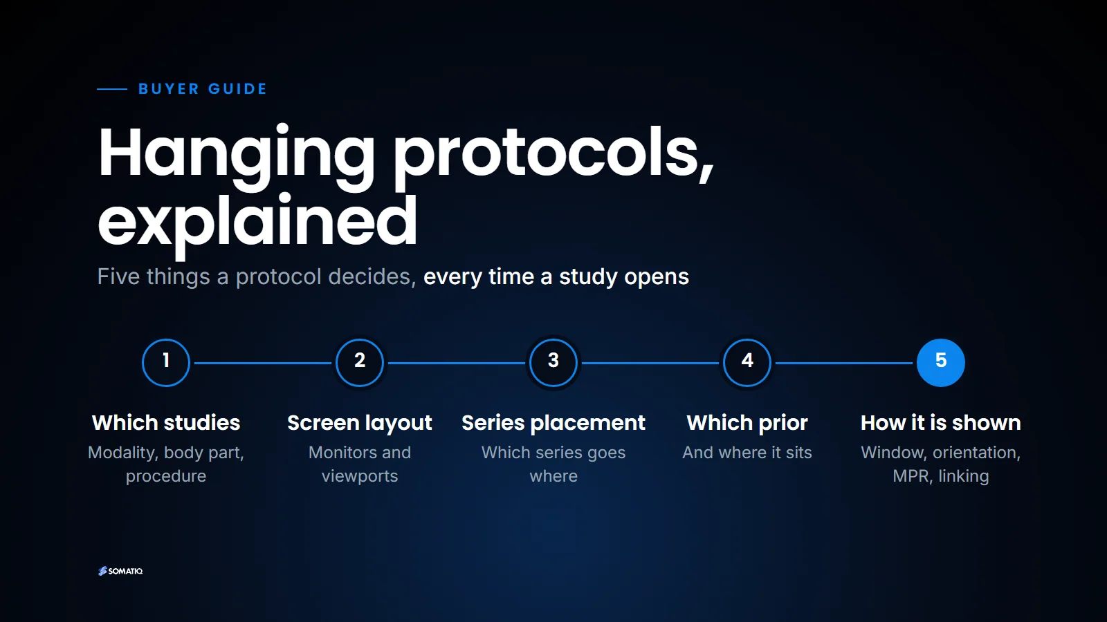
A reasonably complete protocol answers five questions.
Which studies does it apply to? Usually by modality, body part and procedure. "MR brain with contrast" is a different layout from "MR lumbar spine".
What is the screen layout? How many monitors, and how many viewports on each: a 2 x 2 on one screen, a 1 x 2 on another.
Which series goes into which viewport? Axial T2 top left, FLAIR top right, DWI bottom left, post-contrast T1 bottom right, for example.
Which prior, and where? The most recent comparable study, with matching series placed beside or below the current ones.
How is each image shown? Window preset, orientation, zoom, whether MPR or MIP is pre-built, and whether series are linked for synchronised scrolling. For fused studies such as PET/CT, it also decides which viewports show the fusion (see Reading PET/CT in the browser).
The clever part is the matching. The protocol does not know that a series is "axial T2". It has to work that out from the DICOM headers, i.e. from attributes such as Modality, Body Part Examined, Series Description and Image Orientation (Patient). (If those terms are new, our DICOM explainer covers the basics.) The procedure code on the order helps too, which is one more reason a clean order from the RIS or HIS matters downstream.
Why hanging protocols break
Here is the honest reason most radiologists have a love-hate relationship with them. They work beautifully on the studies from your own scanner and fall apart on everything else.
Herman Oosterwijk, the PACS and DICOM educator, put it well in his May 2026 article What's beyond PACS 3.0?. Hanging protocols, he writes, "kind of work… as long as you don't have too many priors" created by another facility or manufacturer that uses different study and series descriptions and body part encoding.
That matches what we see in Indian centres. A few examples of the same thing labelled differently:
| What the radiologist means | What the headers might say |
|---|---|
| Axial T2 brain | "T2_TSE_TRA", "Ax T2 FSE", "T2W_TSE_axial", "t2 tra" |
| Plain CT chest | "CT CHEST PLAIN", "Thorax 1.0 B70f", "Lung 1.25mm", "HRCT" |
| Body part | "CHEST", "THORAX", "LUNG", or left blank |
Now add the realities: a chain with a GE scanner at one branch, a Siemens at another and a refurbished Philips at a third; protocol names typed by different technicians over ten years; a software upgrade on the scanner that silently renamed half the series; priors that came in from an outside centre on a CD. A protocol that matches on Series Description text will miss a good share of these. The radiologist then sees an empty viewport or, worse, the wrong series in the right place.
What the DICOM standard offers
DICOM does have a standard for this. The Hanging Protocol work was added through Supplement 60, final text dated 18 January 2005. It defines:
- a Hanging Protocol IOD in PS3.3, an object that captures a user's or group's viewing preferences for a type of study, with definition, environment and display modules;
- a Hanging Protocol Storage SOP Class and a Hanging Protocol Query/Retrieve Service Class in PS3.4, so protocols can be stored, found and retrieved between systems using C-FIND, C-MOVE and C-GET.
Both are still part of the current standard; they have not been retired. The idea is sensible. A radiologist's protocols could live as objects in the archive and follow them from one workstation to another, or even from one vendor's viewer to another's. In practice, in our experience, very few installed PACS exchange hanging protocols this way. Most keep their protocols in their own proprietary format, so if you change PACS, expect to rebuild them.
The standard also does not solve the naming problem. A DICOM Hanging Protocol object still has to select images by attribute values. If one scanner writes "THORAX" and another writes nothing, the object is no smarter than the vendor's own rules. The advantage of the standard is portability; its disadvantage is that it inherits every inconsistency in the headers it reads.
Practical setup advice
This is what we would tell a centre setting up or cleaning up its hanging protocols.
Start with your top five study types. For most Indian centres, that is something like CT brain, CT chest, MRI brain, MRI lumbar spine and chest X-ray. Get those right before touching anything else; they are most of the volume.
Clean up at the source. Go to each scanner and standardise protocol and series names, with the application specialist if needed. It is tedious work, done once, and it helps everything downstream: hanging protocols, prior matching, AI routing, even billing reports.
Map, do not match strings. Keep a mapping table per scanner: this Series Description from this station means "axial T2". Use more stable attributes where you can, such as modality, orientation from Image Orientation (Patient), slice thickness, contrast flag and sequence parameters, rather than relying only on free-text descriptions.
Decide the prior rule explicitly. Most recent comparable study? Most recent of the same modality within two years? Write it down. A protocol that pulls a five-year-old CT KUB as the "prior" for a CT chest is worse than none.
Build a fallback. When matching fails, the viewer should show the series in a sensible default order, not empty viewports. A radiologist will forgive a fallback layout. They will not forgive a blank screen.
Separate personal from shared. Let the department agree on base protocols, and let individual radiologists save tweaks on top. When a new radiologist joins, they start from the shared set, not from zero.
Re-test after every scanner upgrade. Upgrades rename things. Open five studies from each scanner after any software change and check.
Check from outside the reading room. If your radiologists read from home or from another centre, open studies there too. Monitor count and resolution change the layout that makes sense, and a tablet needs a different layout from a dual-monitor desk.
Where software can help now
Matching on text is the weak point, so this is where smarter software earns its keep. A viewer can learn that a station's "T2W_TSE_axial" is the same thing as another's "Ax T2 FSE" once a radiologist corrects it, and apply that correction everywhere. Herman's article lists hanging protocols among the areas where the industry now expects AI to help, and we agree it is a sensible target. It is a classification problem with a lot of examples and a clear right answer.
In SOMATIQ, the viewer is zero-footprint (see What is a zero-footprint DICOM viewer) and supports custom hanging protocols, along with 3D/MPR and access from mobiles and tablets. Because it runs in the browser, there is no separate workstation to configure at each centre or home office where a radiologist reads; the protocols travel with the user rather than with the machine.
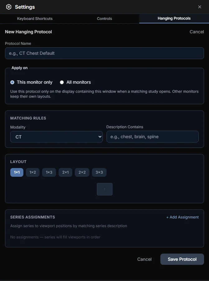
Creating a hanging protocol in the SOMATIQ viewer: name it, say which studies it matches, pick a layout, and assign series to viewports.
So where does that leave a centre that is tired of dragging series around? The practical upshot is that most of the fix happens before the viewer: consistent naming at the scanner, a mapping table for what cannot be renamed, and an explicit prior rule. Once those are in place, any decent viewer's protocols start to behave, and the remaining mismatches are few enough for software to learn from.
Related reading
FAQ
What is a hanging protocol in PACS?
It is a set of rules that decides how a study is laid out on screen when it opens: which series go in which viewport, with which window settings, and which prior is shown beside it.
Why do hanging protocols fail on priors?
Because they match images using DICOM header values such as series descriptions and body part, and different scanners and facilities label the same thing differently. Studies from another centre or vendor often do not match.
Is there a DICOM standard for hanging protocols?
Yes. Supplement 60 (2005) added the Hanging Protocol IOD in PS3.3 and the Hanging Protocol Storage and Query/Retrieve services in PS3.4. They remain in the current standard, though few PACS use them to exchange protocols in practice.
How do I make hanging protocols more reliable?
Standardise series naming at each scanner, keep a mapping table per station, use stable attributes rather than text alone, define the prior rule explicitly and re-test after every scanner upgrade.
Ready to see your workflow on one platform?
We will walk you through the whole workflow, from registration to signed report.